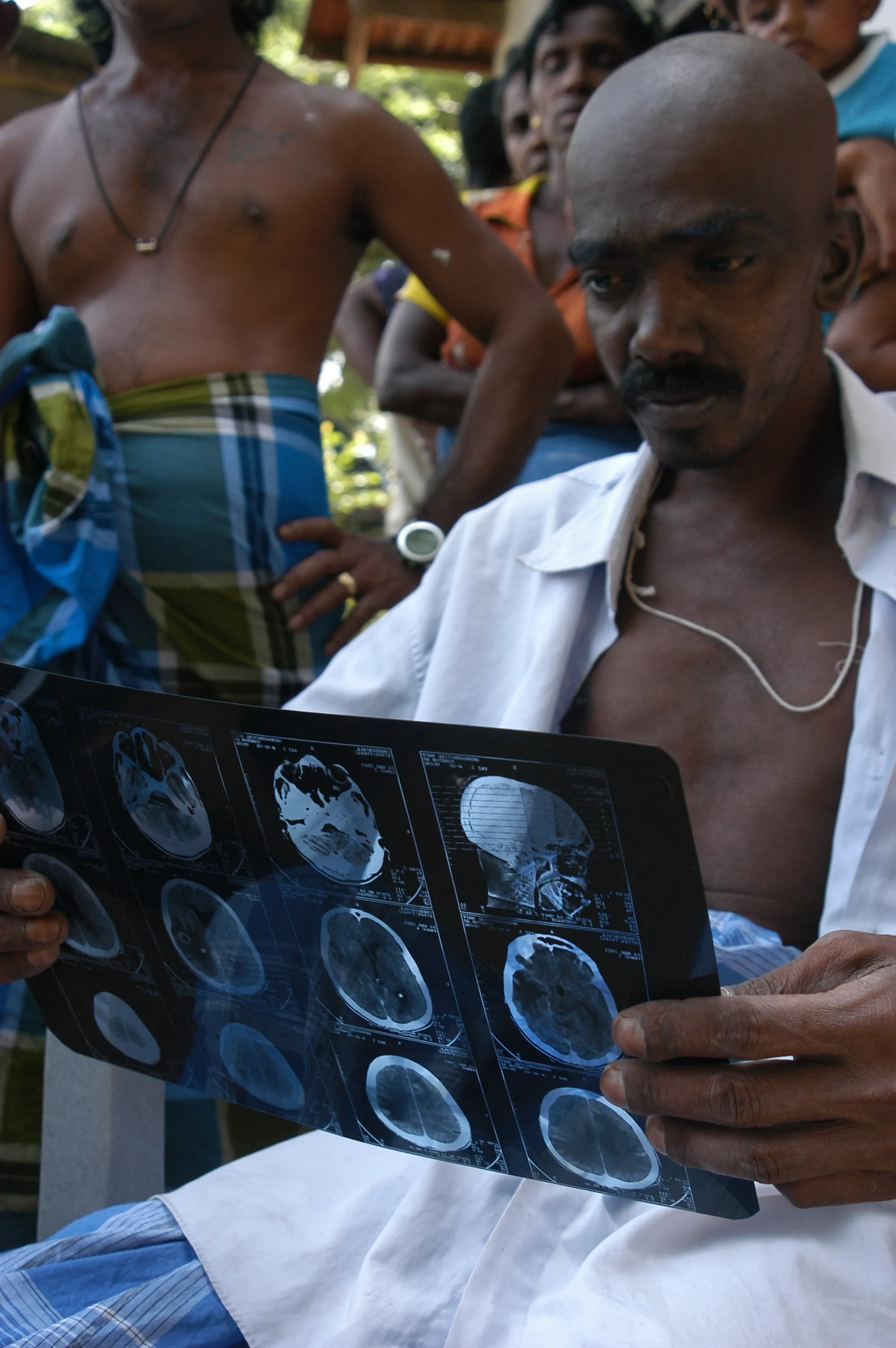
Diagnostic imaging can be divided into two broad categories: those methods that define very precisely anatomical details and those that produce functional or molecular images.
The first method (using CT and MRI) can provide exquisite details on lesion location, size, morphology and structural changes to surrounding tissues, but only delivers limited information as to the tumour’s functioning.
The second method (using PET and SPECT) can give insight into the tumour physiology down to the molecular level, but cannot provide anatomical details.
Combining these two methods enables the integration of anatomy and function in a single approach. The introduction of such “hybrid” imaging has allowed for the characterization of tumours in all stages.
